Endoscopy is the examination of human internal organs using a special high-precision medical device - an endoscope.

An endoscope is a long, flexible tube with an optical system at the end, which projects the image of the examined organ onto a monitor screen. Endoscopy has become most widespread in diagnosing and treating gastrointestinal tract diseases, making a revolutionary breakthrough in gastroenterology and coloproctology.

This method allows not only to visually assess the condition of organs, detect inflammation, bleeding, erosions, but also to identify and prevent the development of oncological diseases. Special endoscope manipulators enable the collection of material for further examination (biopsy) or the removal of polyps.

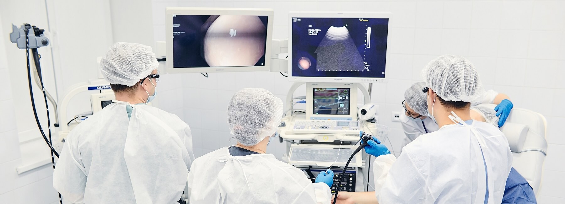
The endoscopy department of the EVIMED Clinic is equipped with the latest expert-class Japanese equipment. All examinations are performed by experienced and highly qualified doctors capable of solving serious diagnostic and therapeutic tasks. This allows us to conduct a full diagnostic complex of gastrointestinal examinations in the shortest possible time and in the most comfortable conditions for the patient.

The processing of endoscopic devices and instruments after examinations is carried out in a specially equipped room, according to sanitary and epidemiological rules and methodological instructions. The storage of endoscopes is carried out in a special cabinet with a bactericidal recirculator, eliminating the possibility of secondary contamination and infection of patients during endoscopic examinations. Strict daily control is maintained over compliance with requirements for processing and disinfection of endoscopic devices and their instruments.

  • EGD (Esophagogastroduodenoscopy)

    Esophagogastroduodenoscopy, or gastroscopy, fibrogastroscopy, EGD, or EGS is a method of visual examination of the upper gastrointestinal tract (esophagus, stomach, duodenum) that allows for the detection of anatomical changes in the upper gastrointestinal tract, a detailed examination of the mucous membrane of the esophagus, stomach, and duodenum, the detection of inflammatory or tumorous pathology, specification of the localization and extent of the process, and also for performing targeted biopsies.

    The most common diseases of the gastrointestinal tract include gastritis, gastroesophageal reflux disease and its complications, duodenitis, peptic ulcer disease of the stomach and duodenum, benign and malignant neoplasms. One of the most important functions of endoscopic examinations is the detection of precancerous changes and early forms of malignant diseases, the risk of which increases with age.

  • Indications for Endoscopic Examination:

    • All men and women aged 40 and older should undergo EGD - an endoscopic examination of the stomach at least once. This is necessary primarily for the prevention of oncological diseases. Referrals for the examination can come from doctors of various specialties;
    • Patients who have been taking medications for a long time, such as hormones, anti-inflammatory drugs, or many cardiological drugs, should undergo endoscopic examination of the stomach. This is necessary to prevent potential complications from long-term use of such medications;
    • Patients with a family history of any oncological diseases should also undergo periodic endoscopic examination;
    • The patient's own desire can also be an indication for endoscopy.
  • Contraindications for Endoscopic Examination

    • Acute inflammatory diseases of the mouth and throat;
    • Acute myocardial infarction;
    • Acute cerebral circulation disorders;
    • Severe congestive heart failure (Stage III);
    • Mental disorders.
  • Indications for Colonoscopy

    • Presence of blood in stool or rectal bleeding;
    • Abdominal pain, especially in the lower abdomen;
    • Changes in stool characteristics (such as the onset of constipation, loose or watery stool);
    • Need for monitoring treatment progress;
    • Detection of hidden blood in the stool;
    • Elevated levels of specific cancer markers in blood tests;
    • Loss of appetite and weight;
    • Familial history of colorectal cancer;
    • Pain along the course of the colon;
    • Sensation of a "foreign body" in the colon;
    • Subfebrile temperature (persistent low-grade fever) of unclear etiology;
    • Anemia (low hemoglobin levels);
    • Suspected tumors or polyps;
    • Age over 45 years (for colon cancer prevention).
  • Conducting Endoscopic Examinations under Sedation

    Conducting endoscopic examinations under sedation (light anesthesia) completely eliminates pain and emotional discomfort for the patient, allowing the doctor to conduct a more thorough examination, fully distend the stomach or intestine using carbon dioxide gas, and closely examine all detected changes.

    Immediately after the examination is completed, you will wake up, and until you fully awaken, you will be under the care of medical staff in the ward.

  • Stomach and Intestinal Polyps

    Stomach and intestinal polyps are benign glandular growths that originate from the epithelial tissue of the stomach and intestinal wall and protrude into the lumen of the hollow organ. Despite their benign nature, they pose several dangers, including:

    • High risk of bleeding from a damaged polyp;
    • Induction of disturbances in the functions of the stomach and intestines (dyspepsia, stasis of contents, motility and evacuation disorders, etc.);
    • Malignant transformation (malignancy).

    Polyps can have various locations and sizes, be single, multiple, or diffuse along the walls of the stomach or intestine (polyposis). The condition often progresses asymptomatically, but even in the absence of complaints in the patient, the recommended course of action upon the accidental discovery of a polyp is its surgical removal.

    Timely diagnosis allows for the removal of potential cancer through a minimally traumatic method, which implies:

    • Minimal risk of complications;
    • Mild postoperative symptoms (pain, weakness, dizziness, etc.);
    • Short recovery period.
  • Advantages of Endoscopic Examinations at the EVIMED Medical Center Include:

    1. State-of-the-art equipment and maximum visualization capabilities.

      Patients often fear endoscopic examinations, recalling old devices. In reality, the innovative equipment of the clinic is designed to minimize discomfort while providing maximum information. We use equipment from the leading Japanese manufacturer Olympus, featuring high-quality optics and thin flexible endoscopes. We also employ NBI, an innovative narrow-band imaging system that allows the doctor to see the smallest changes in the mucous membrane and lesions as small as a few millimeters.

    2. Disinfection and sterilization system.

      Our clinic strictly adheres to hygiene and safety requirements during procedures. Equipment is processed in accordance with sanitary and epidemiological regulations and guidelines. Flexible endoscopes are stored in aseptic conditions in specialized cabinets.

    3. Ability to perform the examination under sedation without pain or discomfort.

    4. Additional diagnostic and therapeutic possibilities.

      Our specialists can also perform tissue sampling for biopsy or carry out therapeutic procedures (removal of growths, stopping bleeding, etc.)

  • Sigmoidoscopy

    Sigmoidoscopy is a method of endoscopic diagnosis that allows for the visual examination of the mucous membrane of the rectum and sigmoid colon.

    Sigmoidoscopy differs from rectosigmoidoscopy in the type of endoscope used. During sigmoidoscopy, a flexible instrument is used, whereas with rectosigmoidoscopy, a rigid one is used. Rectosigmoidoscopy assesses only the rectum and the distal (farther from the hypothetical center) part of the sigmoid colon. Sigmoidoscopy can assess both the rectum and the entire sigmoid colon and, in some cases, the distal part of the large intestine.

    Sigmoidoscopy is often confused with colonoscopy. However, colonoscopy and sigmoidoscopy have specific differences, the main one being the depth of the intestine at which the examination is performed.

    Sigmoidoscopy is often performed when it is not possible to prepare thoroughly for colonoscopy for various reasons. Colonoscopy is recommended when the pathology is located deeper in the intestine than the sigmoid colon.

    Indications for sigmoidoscopy:

    • Pain in the left lower abdomen.
    • Pain and discomfort during defecation.
    • Constipation and narrow ribbon-like stools, indicating a blockage in the rectum and sigmoid colon.
    • Presence of bloody, purulent, or mucous impurities in fecal matter.
    • Positive tests for hidden blood in the stool.
    • Iron-deficiency anemia, often caused by occult intestinal bleeding.
    • Abdominal distension, bloating, rumbling in the lower left abdomen.
    • Diarrhea accompanying inflammation of the rectum (proctitis) and sigmoid colon (sigmoiditis).
    • Suspected familial polyposis, a condition where polyps accumulate in the large intestine. This examination is advisable for individuals with a family history of the condition.
    • Monitoring the condition of the intestines in cases of chronic pathologies.
    • Positive results in blood tests for tumor markers.
    • Fecal incontinence.
    • Monitoring the condition of the intestines in cases of Crohn's disease, a disease characterized by ulceration of tissues.
    • Presence of widespread endometriosis, a condition where endometrial cells from the uterine lining appear in various areas of the body, including the large intestine.

    Sigmoidoscopy allows for the confirmation of conditions such as colitis or cholecystitis. It is also performed after gallbladder removal and as part of preparation for ultrasound examinations. Proctologists use sigmoidoscopy for tissue sampling if laboratory analysis is required. The method has proven effective in detecting signs of cancer, removing foreign objects from the intestines, and monitoring the results of various types of therapy.

  • Preparation for Sigmoidoscopy:

    Usually, about a day before the scheduled procedure, the patient should stop eating. Drinking water is allowed. A few hours before the examination, in this case, it is necessary to cleanse the intestines using an enema.

    In some cases, for example, if there is suspicion of inflammation of the intestinal mucosa, performing an enema is not recommended. This is because its placement can have a negative impact on the condition of the mucous membrane and complicate its visual examination. Detailed instructions on preparing for sigmoidoscopy in this case will be provided by the attending physician.

  • How Does the Sigmoidoscopy Procedure Work?

    The sigmoidoscope has a channel for instruments. Therefore, during the procedure, the doctor can take tissue samples from various sections of the rectum and colon, seal blood vessels, remove polyps, and perform other therapeutic and diagnostic procedures.

    After sigmoidoscopy, some discomfort and mild pain in the area of the rectum and sigmoid colon may be observed for some time, as well as slight traces of blood in the stool. Such a condition does not require any medical intervention.

    How Does the Procedure Work?

    Several days before the procedure, the patient should follow specific recommendations to prepare the body for the examination. The doctor prescribes a diet and a course of cleansing enemas. The patient only drinks water and refrains from eating for a day before sigmoidoscopy. The preparation and its sequence are determined individually, depending on the patient's body characteristics and the likely diagnosis.

    Sigmoidoscopy is performed on a bed with the patient lying on the left side. Initially, the doctor examines the intestine through palpation to ensure there are no clear contraindications. If the patient is male, prostate examination is conducted simultaneously. Afterward, a flexible endoscope with a miniature camera at the end is inserted into the rectum.

    As the sigmoidoscope is advanced into the rectum of the patient, the doctor examines the condition of the mucous membrane, studies polyps, ulcers, fissures, hemorrhoidal nodes, neoplasms, and other pathological areas. If necessary, a tissue sample may be taken for biopsy.

    At the EVIMED Medical Center, this and other endoscopic examinations are conducted by qualified specialists with extensive experience. They know how to minimize patient discomfort during sigmoidoscopy. If the patient complains of painful sensations, the introduction of the endoscope is stopped.

    This method has several advantages over other types of colonoscopic examinations:

    • Compared to colonoscopy, which examines the entire colon, sigmoidoscopy is easier to tolerate and quicker. The procedure can be performed without anesthesia. During colonoscopy, 120-150 cm of the colon is examined, while sigmoidoscopy has a shorter length, up to 60 cm. Furthermore, the preparation before colonoscopy needs to be more thorough to completely empty the colon of fecal matter.
    • Compared to rectosigmoidoscopy, sigmoidoscopy provides the opportunity to examine not only the very bottom (distal) part of the sigmoid colon but the entire sigmoid colon itself.
  • Rectoscopy

    Rectoscopy is a medical examination technique of the rectum using a rectoscope. This device consists of a short tube with an LED light at the end, and it has a mechanism for air insufflation. The tube is inserted into the anal opening, and air is used to inflate the rectum. Rectoscopes in the RMC clinic's endoscopy department are equipped with a fiber optic video system and a device for taking biopsies and removing growths.

    This method helps diagnose:

    • hemorrhoids
    • ulcerative colitis
    • rectal fissures
    • chronic or acute paraproctitis
    • anal fistulas
    • prostate adenoma (in men)
    • rectal endometriosis (in women)
    • polyps and tumors

    Rectoscopy is performed not only for diagnostic purposes. Preventive examination is recommended for men and women over 40 years of age. At this age, the risk of developing proctological diseases that can be successfully treated at early stages increases.

    Indications and Contraindications for Rectoscopy

    Rectoscopic examination is prescribed for patients with complaints of unpleasant symptoms and abnormalities in the functioning of the gastrointestinal tract, including:

    • altered bowel habits
    • defecation problems
    • bleeding
    • inflammatory bowel diseases
    • signs of hemorrhoids
    • mucous or purulent discharge from the rectum
    • suspected growths

    Contraindications to rectoscopy include peritonitis, rectal bleeding, inflammatory and neoplastic processes in the anal area, and acute paraproctitis.

    How Is the Diagnostic Procedure Conducted?

    Two days before the scheduled examination, the patient is advised to reduce physical activity and is given a special diet. To completely cleanse the intestines, two enemas are performed: one the evening before and the other in the morning of the procedure. The use of laxative medications may also be considered.

    The examination is performed using a rectoscope. It is inserted into the rectum to a depth of 20 to 35 cm. The rectoscope consists of a hollow tube with an illumination device and is equipped with an air insufflation system. Optical eyepieces are attached to the device for inspecting the rectal mucosa.

    If necessary, rectoscopy can be combined with biopsy and minimally invasive therapeutic procedures, such as polyp removal and vascular coagulation.

  • Difference Between Rectoscopy and Colonoscopy

    Essentially, both of these methods are not fundamentally different from each other. However, they have one fundamental difference.

    Rectoscopy allows examination of only the rectum and distal parts of the sigmoid colon, while colonoscopy examines the entire colon. The devices themselves differ in the length of their tubes – a fibrocolonoscope can be up to one and a half meters long, while a rectoscope is no more than sixty centimeters.

    Rectoscopy or colonoscopy: which is better?

    These methods of endoscopic examination are not mutually exclusive. In some cases, both of these examinations are prescribed for maximum clarification of the situation. The reason is that the lower part of the rectum is not visible during colonoscopy, and this limitation is compensated for by rectoscopy.

    Gastric and intestinal polyps are benign glandular neoplasms that originate from the epithelial tissue of the stomach and intestinal walls, protruding into the lumen of the hollow organ. Despite their benign nature, they pose several risks, including:

    • high risk of bleeding from a damaged polyp;
    • provocation of gastric and intestinal dysfunction (dyspepsia, content stasis, motility disorders, etc.);
    • malignant transformation (malignancy).
  • Polyps

    Polyps can have various locations and sizes, be single, multiple, or diffusely spread along the walls of the stomach or intestines (polyposis). The condition often progresses asymptomatically, but despite the absence of complaints in the patient, the removal of a detected polyp is recommended.

    Timely diagnosis allows for the removal of potential cancer using a minimally traumatic method, which involves:

    • minimal risk of complications;
    • mild postoperative symptoms (pain, weakness, dizziness, etc.);
    • short recovery period.

    In the EVIMED Medical Center, polyp removal can be performed during colonoscopy and gastroscopy.

  • Fibrobronchoscopy (FBS)

    This is a method of examining the mucous membrane of the tracheobronchial tree, including the trachea, bronchi, and also the larynx.

    Indications for Performing Bronchoscopy

    • Diagnosis of inflammatory and scar processes in the trachea and bronchi;
    • Diagnosis of tumors of the trachea, central and peripheral lung tumors (benign and malignant) detected on X-rays, with the collection of material for cytological or histological examination to differentiate between malignant and benign formations, determine the morphological structure and extent of the malignant process, as well as any associated diseases with a confirmed diagnosis of cancer;
    • Unclear etiology of bronchial stenoses and bronchiectasis - examination of significantly curved bronchi;
    • Inflammatory processes accompanied by segmental and subsegmental atelectasis or infiltrates;
    • Chronic nonspecific bronchial diseases;
    • Chronic suppurative lung diseases (search for bronchi draining lung abscesses);
    • Hemoptysis;
    • Small foreign bodies embedded in subsegmental or more distal bronchi;
    • Disseminated lung diseases;
    • Suspected tuberculosis;
    • Mediastinal diseases;
    • Pleural diseases;
    • Evaluation of the condition of the bronchial stump after surgery;
    • Monitoring the condition of the trachea and bronchi during endotracheal intubation and tracheostomy;
    • Planning and performing various endoscopic surgeries;
    • Change in the character of cough in a smoker + cough for more than 1 month, against the background of intensive therapy.

    Contraindications and Possible Complications of Bronchoscopy

    Absolute contraindications:

    • Agonal condition;
    • Acute stage of myocardial infarction;
    • Acute stage of stroke;
    • Asthmatic status;

    Relative contraindications:

    • Fever unrelated to a lung process is recommended to postpone the procedure for several days;
    • A patient's condition where bronchoscopy does not provide a solution;
    • Laryngeal stenosis (not a contraindication for fibrobronchoscopy through an intubation tube or tracheostomy);
    • Intolerance to anesthetics (the examination is performed under anesthesia);
    • Severely compromised general patient condition;
    • Absence of signed informed consent from the patient for the procedure (examination);
    • Local changes (significant deformation of the tracheobronchial tree).

    Preparation for the Procedure

    Usually, the procedure is performed in the morning. Food intake is allowed at least 8 hours before the procedure, as bronchoscopy is done on an empty stomach. This is necessary to avoid food residues entering the respiratory tract.

    If the patient is taking medication, they should consult with a doctor about the medication schedule one day before the examination. On the day of bronchoscopy, drinking water and smoking are not allowed.

    Anesthesia for Endoscopic Examinations

    Local anesthesia is used for endoscopic examinations of the upper respiratory tract (nasopharyngoscopy, laryngoscopy, bronchoscopy). If necessary and upon the patient's request, all endoscopic examinations can be performed under sedation (anesthesia).

    How Bronchoscopy Is Performed

    The procedure is performed with the patient sitting or lying down. The doctor sprays anesthetic on the mucous membranes of the nasal cavity, oropharynx, and larynx. Anesthesia not only helps minimize pain but also suppresses coughing. When the medications start to take effect, the patient experiences numbness of the throat and tongue. Subsequently, the endoscope is inserted into the respiratory tract through the nasal or oral cavity. The procedure is painless. Since the diameter of the bronchi is much larger than that of the bronchoscope, there is no discomfort during breathing. The doctor then examines the condition of the bronchi. If necessary, tissue samples are taken for examination (biopsy).

    In the EVIMED center, the doctor can perform the following during bronchoscopy:

    • Lavage and sanitation of the bronchial tree
    • Biopsy
    • Restoration of the bronchial lumen
    • Stenting of the bronchial lumen

    These procedures are performed after a prior consultation with the doctor.

    After the procedure, the patient is provided with video material of the performed manipulation, and a consultation with a specialist is conducted, including recommendations and planning of further treatment or observation.

    If material is taken for histological or cytological examination, the results are issued within 5-7 working days.

    Subsequent Recovery after Bronchoscopy

    After a routine diagnostic procedure, it is advisable to wait for some time before eating until the local anesthetic effect in the throat completely disappears (30-40 minutes). After general anesthesia, nervous system recovery occurs within a day, and during this time, it is not recommended to drive a car or engage in activities that require attention.

  • Helicobacter pylori

    Helicobacter pylori is a widely prevalent gastric infection agent that causes pathologies such as gastritis, peptic ulcer disease, adenocarcinoma, and low-grade lymphoma of the stomach. The diagnosis is established based on a breath test, stool antigen analysis, or samples obtained during endoscopic biopsy.

    The most reliable diagnosis involves examining material obtained during endoscopic procedures. The collected biopsy sample (a piece of the mucous membrane) is placed in a special reagent, and when evaluating changes in parameters, a specialist provides a conclusion of a positive or negative result.

    The breath test is a non-invasive method of examination. It is safe for small children and pregnant women. It can be used for primary diagnosis of Helicobacter pylori as well as for monitoring the progress of anti-Helicobacter therapy and assessing the effectiveness of previously conducted treatment. The test system has a sensitivity of 95%, and specificity (accuracy of the examination) of 97%. This method is non-invasive, fast, cost-effective, and painless. You receive the results immediately after completing the test.

    At the EVIMED medical center, you can undergo an analysis for Helicobacter pylori. You can also schedule an appointment with a therapist or gastroenterologist to consult about any symptoms that concern you.

  • Endoscopic Biopsy

    Biopsy is a diagnostic procedure performed to obtain a tissue sample (biopsy specimen). Biopsies are typically taken from suspicious abnormal formations. Examples include tumors, non-healing wounds, polyps, and moles. The technique for conducting the procedure and the necessary instruments are determined based on the location of the biopsy.

    The tissue samples obtained through biopsy are sent to a specialized laboratory where a histological analysis is performed. This analysis is based on the fact that all cells in the body have a characteristic structure depending on the tissue to which they belong. When malignancy occurs, the cell's internal structure is disrupted, making it unlike neighboring cells. These changes are usually significant enough to be visible under a regular microscope.

    The primary goal of biopsy is to accurately determine whether a new formation is malignant or benign.

    Another important piece of information that the examination can provide is how effective the treatment of oncology is in each specific case.

    At the EVIMED medical center, various types of biopsies can be performed. We have highly qualified doctors and use modern equipment.

Specialists in the Field

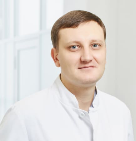
Andrey Mykhailovych Mikita

Physician - Endoscopist

Vladimir Nikolaevich Korolev

Deputy Director for Medical Affairs
Physician - Endoscopist